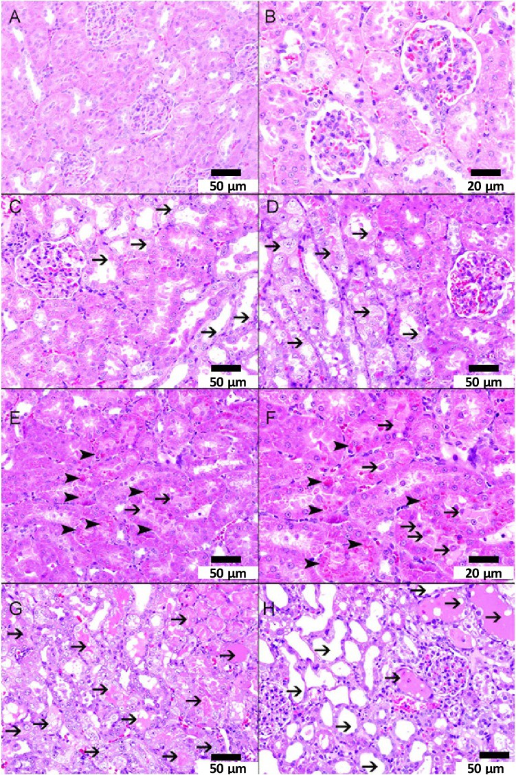
Fig. 5. A-H: representative light microscopy sections of H and E stained kidney tissues of Wistar rats intratracheally instilled with either saline (control) or cerium oxide nanoparticles (CeO2 NPs) with or without cisplatin (CP) administration. A and B: representative kidney sections obtained from saline-treated rats showing normal kidney architecture and histology. There was no tubular injury (0%, n=6) in the examined section. C and D: representative kidney sections obtained from CP + saline-treated rats showing the presence of acute tubular injury in 37.50 ± 2.49 % (n=6) of examined tissue areas (thin arrows) (score 2). E and F: representative kidney sections obtained from CeO2 NPs-treated rats showing the presence of focal acute tubular injury in 7.37 ± 0.94% (n=6) of examined tissue areas with intracytoplasmic aggregates of giant lysosomes within proximal tubular cells (arrowhead), and epithelial cells falling within the lumen of tubular cells (thin arrow) (score 1). G and H: representative kidney sections obtained from CP + CeO2 NPs-treated rats showing the presence higher percentage of acute tubular injury when compared with either CP + saline group or CeO2 NPs group. There is acute tubular injury (thin arrow) involving 58.63 ± 6.27% (n=6) of examined areas (score 3).